Femoroacetabular impingement (FAI) occurs when extra bone ‘spurs’ grow on one or both of the bones in the hip joint, which restricts movement and can cause damage such as tears or Arthritis (degradation of the cartilage).
The three types of FAI are determined by the location of the spurs:
Hip Impingement, or FAI, begins during childhood growth. There is little that can be done to prevent the formation of spurs growing on the bone.
The actual number of people living with FAI is unknown as many live unaffected, active lives. Hip Impingement becomes a problem when these spurs damage cartilage or other parts of the joint.
Certain health conditions and activity could increase the risk of developing FAI. Those engaging in more physical activity may experience more wear and tear in the joint. Conditions such as Legg-Calve-Perthes which affects blood flow into the hip can impact the health inside the joint, or the rare Coxa Vara condition where hip bones grow at differing rates during childhood can cause FAI.
Physical activity involving repetition of flexing the joint past the typical range of motion, such as dance, football and golf, may increase a person’s risk of developing symptoms.
Alignment problems can also prevent smooth movement and lead to impingement or arthritis.
Early FAI does not usually cause symptoms, but as the condition develops symptoms worsen:
The hip is a ball-and-socket joint, which is one of only two in the body (the other is in the shoulder). This type of joint offers maximum mobility and rotation.
The ball is the spherically shaped end of the femur (thigh bone), and the socket is the acetabulum – the cup-shaped area on the side of the pelvis.
Between these two bones, there is a layer of cartilage to ensure smooth gliding of the bones within the joint. It also includes a rim of fibro cartilage (acetabular labrum), which encircles the head of the femur.
Surrounding the joint there is a complicated set of ligaments for stabilization and a short ligament deep inside the joint to hold the ball in the socket.

The first option for mild case relief is nonoperative, and could include:
When other therapies are not appropriate or have not succeeded, surgery may be considered. The aim of which is to firstly repair any damage in the hip joint and secondly reshape the bone to remove spurs.
Where possible, surgery will be done arthroscopically (minor key hole incision with miniature camera and tools). However, some cases may require open surgery, involving a larger incision.
For some, especially in severe cases, this surgery may not be able to offer a complete recovery as more growth may develop in future, requiring future treatment.
Despite this, surgery is currently the best treatment for painful Hip Impingement and will ease symptoms in most cases.
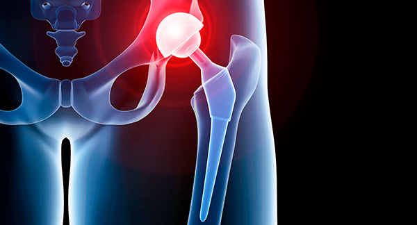
The last line in treatment options is hip replacement surgery. This procedure removes the damaged joint and replaces the ball and socket with prosthetics.

If you are looking to book an appointment, please call us on